Cleveland Clinic Lerner Research Institute Logo
  • About
  • Science
    • Laboratories
    • Office of Research Development
    • Clinical & Translational Research
      Participating in Research
    • Departments
      Biomedical Engineering Cancer Biology Cardiovascular & Metabolic Sciences Florida Research & Innovation Center Genomic Medicine Immunotherapy & Precision Immuno-Oncology
      Infection Biology Inflammation & Immunity Neurosciences Ophthalmic Research Quantitative Health Sciences Translational Hematology & Oncology Research
    • Centers & Programs
      Advanced Musculoskeletal Imaging Angiogenesis Center Cardiovascular Diagnostics & Prevention Computational Life Sciences Consortium for Pain Genitourinary Malignancies Research Genome Center
      Microbiome & Human Health Musculoskeletal Research Northern Ohio Alcohol Center Pathogen & Human Health Research Populations Health Research Quantitative Metabolic Research Therapeutics Discovery
  • Core Services
    • Ohio
      3D Printing Bioimage AnalysisBioRobotics & Mechanical Testing Cell Culture Cleveland Clinic BioRepository Computational Oncology Platform Computing Services Discovery Lab Electron Microscopy Electronics Engineering
      Flow CytometryGenomic Medicine Institute Biorepository Genomics Glassware Histology Hybridoma Immunohistochemistry Immunomonitoring Lab Instrument Refurbishing & Repair Laboratory Diagnostic
      Lerner Research Institute BioRepository Light MicroscopyMechanical Prototyping Microbial Culturing & Engineering Microbial Sequencing & Analytics Resources Media Preparation Molecular Biotechnology Nitinol Polymer Proteomics & Metabolomics Therapeutics Discovery
    • Florida
      Bioinformatics
      Flow Cytometry
      Imaging
  • Education & Training
    • Graduate Programs Molecular Medicine PhD Program Postdoctoral Program
      Research Intensive Summer Experience (RISE) Undergraduate & High School Programs
  • News
  • Careers
    • Faculty Positions Research Associate & Project Staff Postdoctoral Positions Technical & Administrative Engagement & Belonging
  • Donate
  • Contact
  • About
  • Science
    • Scientific Programs
    • Laboratories
    • Office of Research Development
    • Clinical & Translational Research
      Participating in Research
    • Departments
      Biomedical Engineering Cancer Biology Cardiovascular & Metabolic Sciences Florida Research & Innovation Center Genomic Medicine Immunotherapy & Precision Immuno-Oncology
      Infection Biology Inflammation & Immunity Neurosciences Ophthalmic Research Quantitative Health Sciences Translational Hematology & Oncology Research
    • Centers & Programs
      Advanced Musculoskeletal Imaging Angiogenesis Center Cardiovascular Diagnostics & Prevention Computational Life Sciences Consortium for Pain Genitourinary Malignancies Research Genome Center
      Microbiome & Human Health Musculoskeletal Research Northern Ohio Alcohol Center Pathogen & Human Health Research Populations Health Research Quantitative Metabolic Research Therapeutics Discovery
  • Core Services
    • All Cores
    • Ohio
      3D Printing Bioimage Analysis BioRobotics & Mechanical Testing Cell Culture Cleveland Clinic BioRepository Computational Oncology Platform Computing Services Discovery Lab Electron Microscopy Electronics Engineering >
      Flow CytometryGenomic Medicine Institute BiorepositoryGenomics Glassware Histology Hybridoma Immunohistochemistry Immunomonitoring Lab Instrument Refurbishing & Repair Laboratory Diagnostic
      Lerner Research Institute BioRepository Light MicroscopyMechanical Prototyping Microbial Culturing & Engineering Microbial Sequencing & Analytics Resources Media Preparation Molecular Biotechnology Nitinol Polymer Proteomics & Metabolomics Therapeutics Discovery
    • Florida
      Bioinformatics
      Flow Cytometry
      Imaging
  • Education & Training
    • Research Education & Training Center
    • Graduate Programs Molecular Medicine PhD Program Postdoctoral Program
      Research Intensive Summer Experience (RISE) Undergraduate & High School Programs
  • News
  • Careers
    • Faculty Positions Research Associate & Project Staff Postdoctoral Positions Technical & AdministrativeEngagement & Belonging
  • Donate
  • Contact
  • Search

Research News

❮News Looking inside the "black box" of neural activity to aid stroke research

08/26/2024

Looking inside the "black box" of neural activity to aid stroke research

Dr. Hod Dana partners with Drs. Ken Baker and Andre Machado to help understand neural activity behind deep brain stimulation for stroke.

Dr. Hod Dana examines machines in his lab. He is mainly in darkness, but backlit.
Dr. Hod Dana's lab develops advanced imaging technologies for tracking neural activity, including calcium imaging.

Neural activity, or how electric currents move between brain cells called neurons, influences everything from longevity to neurodegenerative disease. Researchers have long attempted to see inside the metaphorical “black box” of the brain to understand neurological disease.

Hod Dana, PhD, has built a research program around capturing the lightning-fast flashes of neural activity that are key to everyday activities. His next step is using novel advanced imaging and microscopy techniques to decode the successful results of a phase I deep-brain stimulation (DBS) trial for ischemic stroke.

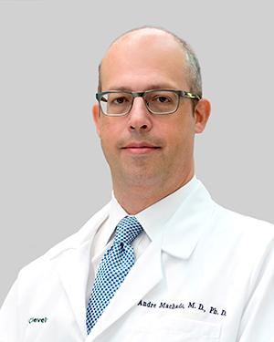
Cleveland Clinic’s Andre Machado, MD, and Ken Baker, PhD, developed a DBS treatment for patients recovering from stroke. The treatment sends gentle electrical currents to a specific part of the brain that controls fine motor movements. A $3.2 million National Institutes of Health grant will fund a collaborative investigation by Drs. Dana, Baker and Machado into what made the DBS trial successful.

Dr. Baker says although the team has a great deal of preclinical and clinical data to support the treatment’s therapeutic potential, they have limited ways to see the treatment’s effects on the neuronal level.

“The work with Dr. Dana’s group will enable us to not only further understand how stimulating the cerebellum alters cerebral cortical activity, but more importantly, allow us to visualize changes in those effects as a function of how we deliver stimulation,” Dr. Baker says. “The latter may be crucial to helping us to further optimize and enhance the effects as we shift towards a more patient-specific approach to delivering therapy.”

What can neural activity tell us about stroke research?

Imaging brain activity is not an easy task. Scientists often can only see a small fraction of the brain at a time because of how quickly electrical signals move throughout neurons. Existing monitoring methods are also restrictive, requiring headpieces that limit normal activity.

During a stroke, blood clots cause damage to the brain and cause patients to lose motor skills like walking or talking. That damage affects neural activity, reshaping how the brain completes certain tasks. The concept behind DBS for stroke is that the mild electrical currents would restore neural activity in those areas of the brain, giving the patient the ability to recover. DBS is precise; researchers need to determine exactly which of the brain’s many electrical processes were affected by the stroke within a complex network of neurons.

Dr. Machado and Baker designed their DBS therapy based on decades of preclinical research to determine the right pieces of the brain to target. With Dr. Dana, the team hopes to more precisely map how stroke affects neural activity and why the new DBS treatment works. Having access to this data will allow the investigators to refine the existing treatment, adjusting the strength and frequency of their electrical signals. This includes the prospect of adapting DBS parameters in real time to each patient, maximizing the potential of small changes to brain networks that can result in motor skills improvements.

Dr. Dana’s imaging techniques are built to capture brain activity as it moves among neurons. His team works on calcium imaging techniques in preclinical models, marking neural activity by marking where calcium levels are shifting. The technique allows images to show activity in multiple neurons simultaneously, creating comprehensive maps of brain activity.

This advanced imaging will provide insight into why some patients in the trial did not respond to DBS and point to new strategies for making the treatment effective.

“I’ve had this project in mind since I joined Cleveland Clinic seven years ago,” says Dr. Dana. “The chance for close collaboration to apply this technology and potentially change people’s lives is incredibly rewarding. To see techniques and devices we’ve ideated, built and refined reach this stage and solve problems is exciting."

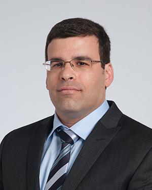
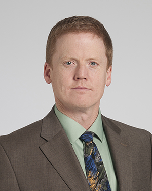
Featured Experts
Hod Dana Headshot
Hod
Dana, PhD
Ken Baker Headshot
Ken
Baker, PhD
Andre Machado Headshot
Andre
Machado, MD, PhD
News Category
Related News
Cleveland Clinic Researchers Awarded $2.5M NIH Grant to Enhance DBS for Stroke RecoveryCleveland Clinic study shows deep brain stimulation encouraging for stroke patientsDepartment of Defense Awards $2.5M for Improving Spinal Cord Injury in Rehabilitation

Research areas

Neurosciences

Want To Support Ground-Breaking Research at Cleveland Clinic?

Discover how you can help Cleveland Clinic save lives and continue to lead the transformation of healthcare.

Give to Cleveland Clinic
Subscribe to get the latest research news in your inbox.

About Lerner

About Us Careers Contact Us Donate People Directory

Science

Clinical & Translational Research Core Services Departments, Centers & Programs Laboratories Research News

Education & Training

Graduate Programs Molecular Medicine PhD Program Postdoctoral Program RISE Program Undergraduate & High School Programs

Site Information & Policies

Search Site Site Map Privacy Policy Social Media Policy

9500 Euclid Avenue, Cleveland, Ohio 44195 | © 2025 Lerner Research Institute